
Lesson 2 - Yeast Morphology and Evaluation
This lesson explores what yeast cells are, how they grow and multiply, what internal organelles they contain, and the functions that these organelles perform. This knowledge will equip us well, so that we can examine some of the practical methods that we use to characterise and evaluate yeast in the brewery.
Yeast Cell Structure and Function
Yeast cells can carry out most of the same functions as cells of higher organisms, including us; however, yeast cells have their own unique set of features, which are not found in any other organisms. These features are described here.
Yeast cells come in a variety of shapes, but are mostly spherical and slightly flattened:
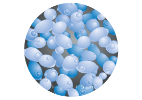
Figure: Scanning electromicrograph of cells of S. cerevisiae
Yeast cells are quite small, usually in the range of 5-10 µM in diameter (1 µM = 1 millionth of a metre); however, they are much bigger than most bacteria (typically, 0.5-2.0 µM). We can see them quite easily under a good quality light microscope. We will expand upon this when we explore the various tests used to assess the quantity and quality of yeast cells using a light microscope.
If we wish to investigate the cells’ internal structures, we will need a much more powerful microscope, alongside special treatments. This will allow us to see a number of sub-cellular organelles. An idealised image of a yeast cell’s sub-cellular structure is shown in the figure below:
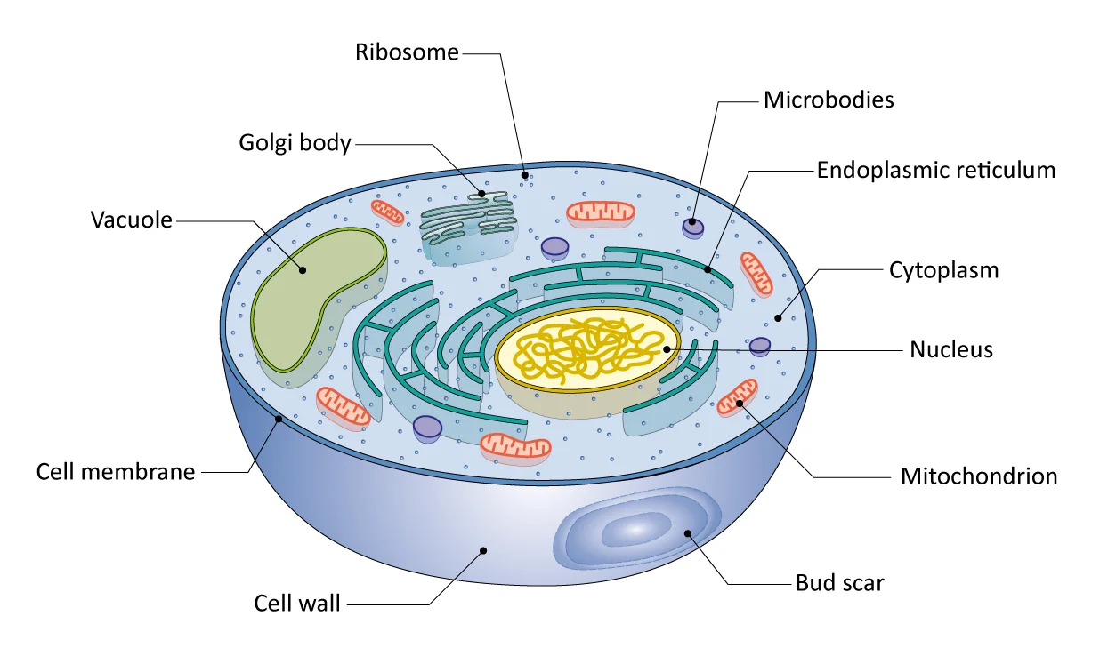
Figure: The main features of a yeast cell
Even using the best light microscope, it is not possible to see much of the detail that we can see in the figure. At best, you should be able to distinguish the vacuoles, but little more than this unless you use specific stains. Most of the organelles shown would require special pre-treatments and an electron microscope in order to see any detail. Each of these organelles has specific functions.
The cellular constituents shown in the figure above are explained in more detail in the appendix at the end of this lesson.
The endosymbiotic theory states that organelles such as mitochondria were once more primitive, free-living, independent organisms. The evidence for this is that mitochondria and other organelles, such as chloroplasts in plants, contain their own DNA where the photosynthetic apparatus is located. Many of their other features are similar to bacterial cells. It is thought that these primitive cells invaded others, originally probably as pathogens, but then gradually adapted to co-exist with the host cell in a symbiotic association. The free-living proto-organelles evolved into prokaryotic cells such as bacteria; the symbiotic associations became the eukaryotic cells.