
Lesson 8 - Yeast Management
This lesson will explore the methods that are used to measure yeast’s concentration and to assess its health. We will then go on to describe how this knowledge underpins the methods used to store and handle yeast in the brewery.
Point of Reflection – Reconvene
Did you think it is a bit more complicated than just describing a viable cell as one that is alive?
We have to think about how we measure ‘life’ in this context. If it were human life, we might look for something simple like a detectable pulse or breathing. If we had the appropriate equipment, we could go for something more complicated, like brain activity. Whatever we choose, we need a measure that we consider is evidence of life.
In the context of yeast and brewing, we have to do the same thing. In this case, we measure something that relates to yeast structure or metabolic activity.
-
The most obvious measure is the ability to grow. We can look at cells suspended in a nutrient medium under a microscope and see if they can bud. Alternatively, we could see if a single cell forms a colony on an agar plate.
-
If the cells were resting and not in a position to grow, we could look for evidence of metabolic activity such as can they take up nutrients and make products of metabolism such as CO2.
All of these approaches are valid and are evidence of something that living cells show.
Is this enough?
Usually, we need a rapid method; we might have a batch of wort ready to be pitched and so would not have the time needed to get a colony count. Assessing metabolic activity can be quick, but it is only applied to bulk populations. Therefore, it is not easy to relate to the viability of individual cells.
The key point is that not all methods give the same answer. A cell might not be able to bud and go onto form a colony on a plate, but it might still be able to take up wort nutrients. Returning to our example on humans, it would be a strange measure of viability if it was based just on the ability to have children!
In brewing, we have to compromise. We want a quick, easy and preferably inexpensive method. The technique that is most often used is a viability stain. There are lots of these stains, as we will go on to discuss. Most rely on viable cells having an intact and functional cell membrane, and the reverse for dead cells.
We would probably get a different result if we used a different method of measuring viability. We accept these shortcomings, but as long as the same method is always used, and the results are reproducible, we can live with the error.
Measuring viability
We need to be able to measure the viability of yeast when it is used in processes dependent on yeast metabolic activity, such as primary fermentation or secondary fermentation in cask or bottle conditioning or classical lagering.
In these processes, we need to know how many viable cells we are dealing with. Just as important is that viability is a good measure of yeast health. If most of the cells are dead, it is a fair guess that the living ones are not in a very happy state.
Colony counts
The classical method is to take a diluted sample of yeast, containing a known number of cells, and plate it out onto an agar plate of nutrient medium to separate the cells. After incubation for two to three days, the viable cells (those able to proliferate) form colonies.
If we count the colonies (called colony forming units, cfu) and compare this figure with the total cell count in the original suspension, we can calculate the viability.
align="center">Viability = Number of cfus / total cell count x 100The method works fine but is too slow for routine use in a brewery. We can make it a bit quicker by using a microscope slide overlayered with a thin film of agar nutrient medium. These are inoculated to give single cells, as before, then examined for the formation of micro-colonies. This approach reduces the time to around twenty-four hours.
Viability strains
Yeast viability is most often measured in brewing using a group of biological dyes that differentially stain viable and non-viable cells.
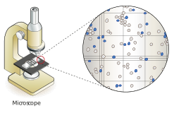
Figure: Yeast cells stained with methylene blue, dead cells stain blue; live cells are unstained and depending on the quality of the microscope used are transparent with little colour and with darker borders
Cells are stained and then examined under a microscope.
These vital stains work in one of two ways:
-
Based on membrane integrity, the stain is not able to enter live cells (with an intact membrane), and so only dead cells (disrupted membrane) are stained.
-
Enters all cells, but the metabolic activity of viable cells causes a colour change; or, viable cells can pump the stain back out. Dead cells with no metabolic activity do not have an effect on the stain.
The most commonly used viability stain for routine brewery testing is methylene blue or the closely related methylene violet. Viable cells are thought to take up stain and change the dye to its colourless leuco form. Dead cells cannot change the dye’s colour. To undertake this test we only need a bright field microscope.
Many of the dyes are fluorescent and need a special microscope capable of dealing with them. As a group, these stains are more expensive than bright field dyes but give much better discrimination, so much so, that they are often used in automatic cell counters where electronic image analysis replaces the human operator (to be discussed later in this lesson). Several fluorescent stains are proprietary.
A few examples of viability strains are shown in the following table:
Table: Examples of viability stains
| Stain | Comments |
| Methylene blue | Dead cells blue, viable cells colourless |
| Propidium iodide |
Enters dead cells only and binds to DNA giving red fluorescence |
| MgANS | Enters dead cells only giving green fluorescence |
| FUN1™ | Enters all cells; dead cells fluoresce green, viable cells develop red fluorescence in internal structures |
| Oxonol | Enters cells with compromised membrane potential giving green fluorescence |
| Fluorescein diacetate | Enters all cells and is modified by active esterase to give a green fluorescent product. These leak from dead cells with permeable membranes and these are not stained |
Many other viability stains are in common use. More information can be found in the following publications:
Kwolek-Mirek, M. & Zadrog-Teiza, R. (2014). Comparison of methods used for assessing the viability and vitality of yeast cells. FEMS Yeast Research, 14, 1068 – 1079.