
Lesson 2 - Mashing Biochemistry
This lesson deals with the biochemical aspects of mashing. We will meet the main substrates and the enzymes that break them down. We will learn what enzymes are, how they operate and how they are affected by the environment of the mash. Finally, we will cover how starch is gelatinised, liquefied and saccharified to make a wort with the desired sugar spectrum.
Protein
We now know a great deal more about the biochemistry of starch and its importance in mashing. Let’s gain the equivalent knowledge about protein.
Like starch, proteins are polymers of simpler molecules. While starch is made up of glucose residues, proteins are made up of amino acid residues. Below is the representation of an amino acid. R denotes a side chain that is specific to the particular amino acid, of which 20 are found in plants.
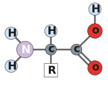
Figure: Amino acid
The protein equivalent to the glycosidic bond is the peptide bond, and like the glycosidic bond, it is formed with the loss of a water molecule. The figure below shows the formation of a peptide bond.
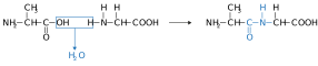
Figure: The formation of a peptide bond
Biochemical names relating to proteins
Peptides are short chains of amino acids linked by peptide bonds. The word peptide comes from the Greek word ‘peptós’, meaning digested. Chains of two and three amino acid residues are called di and tri peptides, while chains up to ten are called oligopeptides. We can see some similarities with carbohydrate naming here. A polypeptide is a continuous chain greater than ten (sometimes greater than 15) amino acid residues. The figure below shows a peptide.
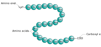
Figure: Peptide
Like chains of glucose residues, chains of amino acid residues have two distinct ends. The -NH2 group, which hasn’t been converted into a peptide link, is shown at the left-hand end and is called the N-terminus (or amino end). The unchanged -COOH group is shown at the right-hand end and is called the C-terminus (or carboxyl end). It is important to distinguish between these ends as they influence the enzymes that catalyse their break down during mashing. More about this later.
Proteins are much more complex than starch and contain nitrogen and sulphur as well as carbon, hydrogen, and oxygen.
To be considered a protein, a molecule usually needs to contain more than 50 amino acid residues. Proteins vary in size from tens to several thousand amino acid residues.
The largest known protein is called Titin and is made up of 34,351 amino acids residues. Bet you are glad you’re a brewer and not a biochemist counting amino acids!
Proteins have primary, secondary, tertiary, and quaternary structures. These are:
-
Primary – the sequence of amino acids held in place by peptide bonds.
-
Secondary – the three-dimensional form of the backbone of the protein. Common secondary structures include the ɑ-helix and the β-strand or sheet. This is held in place by hydrogen bonds.
-
Tertiary – the spatial arrangements of the components of the secondary structure. This is held in place by non-covalent interactions (hydrogen bonding, ionic interactions, van der Waals forces, and hydrophobic packing), and also disulphide bonds and metal ion coordination.
-
Quaternary – the number and arrangement of protein subunits (individual proteins making up the whole protein) often in association with other molecules like metals and nucleic acids.
The figure below shows primary, secondary, tertiary, and quaternary protein structures.
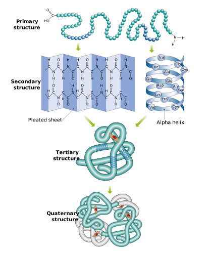
Figure: Primary, secondary, tertiary, and quaternary protein structures.
The link below provides some more information on protein structures.
https://www.ebi.ac.uk/training/online/courses/biomacromolecular-structures/proteins/
Proteins have a diverse range of roles in the biological world, from storage through to enabling animals to get around. This is made possible by these complex and, in some cases, ornate structures.
Proteins are classified according to structure, function, location and/or properties. Many proteins are associated with other compounds and include them in their quaternary structure. Examples include:
-
Carbohydrates (glycoproteins)
-
Lipids (lipoproteins)
-
Metal ions (metalloproteins)
Most of the proteins we need to break down during mashing are relatively simple in structure because they are stores of amino acids that are used to build a new plant. The most important proteins in mashing are the enzymes we use to catalyse the hydrolysis of our mashing substrates. Here, for example, is an artist's impression of the structure of α-amylase which is a metalloprotein incorporating calcium. You can see the calcium ion as the yellowish blob towards the top.
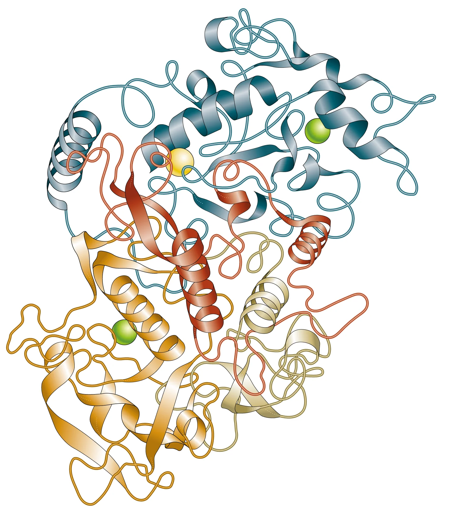
Figure: α-Amylase
We will cover enzymes in depth later in the lesson. Next, we go back to carbohydrates when we discuss non-starch polysaccharides (NSPs).